
Photoacoustic Imaging: Principles and Laser Source Selection
I. Photoacoustic Effect
When a material is irradiated by periodically-modulated or pulsed laser light, it absorbs light energy and undergoes non-radiative relaxation. The absorbed light energy is converted into thermal energy, giving rise to periodic local temperature rise and fall. This triggers thermal expansion and contraction of the material, generating elastic stress that propagates outwards as acoustic waves. This phenomenon is known as the photoacoustic effect.
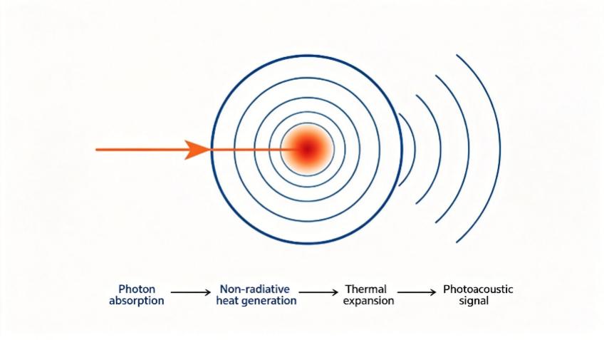
(Schematic diagram of the photoacoustic-effect principle: Photon absorption → Non-radiative heat generation → Thermal expansion → Photoacoustic signal)
The photoacoustic effect was first discovered by Bell in 1880. In his experiments, Bell found that intermittent light beams shone on light-absorbing solid materials generated acoustic waves at the same frequency as the light modulation. This finding laid the foundation for photoacoustic imaging. For nearly a century afterwards, the photoacoustic effect was mainly applied in spectroscopic analysis, which gradually developed into photoacoustic spectroscopy. In the late 20th century, driven by rapid advances in pulsed-laser technology, high-sensitivity ultrasonic detectors and image-reconstruction algorithms, photoacoustic-imaging technology based on the photoacoustic effect achieved extensive development.
II. Photoacoustic Imaging
Photoacoustic imaging is a biomedical-imaging technique built upon the photoacoustic effect. It is a non-invasive, non-ion-radiation detection modality。Working principle: Biological tissue is illuminated by pulsed laser. The tissue absorbs part of the incident light energy; light-energy deposition causes a slight local temperature increase and induces rapid thermo-elastic expansion of tissue. When thermal- and stress-constraint conditions are satisfied, instantaneous thermal expansion generates pressure waves, namely photoacoustic signals. These signals propagate outward in the form of ultrasound waves, are collected by high-sensitivity ultrasonic transducers, and processed via image-reconstruction algorithms to inversely calculate the internal light-absorption distribution of biological tissue, finally producing photoacoustic images.
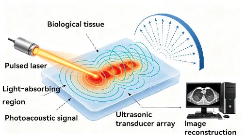
Schematic Diagram of Photoacoustic Imaging Principle
Different biological components differ in light-absorption capacity. For instance, melanin absorbs light far more strongly than adipose tissue under identical conditions. Even the same tissue exhibits variable absorption under laser light of different wavelengths. Leveraging this property, photoacoustic imaging can quantitatively resolve functional parameters such as blood oxygen saturation, reveal tissue composition and metabolic status, and perform functional-level imaging detection.
Photoacoustic signals carry the intrinsic light-absorption information of tissue. This imaging technique combines the high contrast of optical imaging and the large penetration depth of ultrasound imaging. It breaks the ~1 mm imaging-depth limit of conventional optical imaging. Under specific experimental conditions, its imaging depth can reach approximately 70 mm while delivering tissue images with good resolution and contrast. As a fast-growing non-destructive imaging technique in recent years, its spatial resolution spans from micrometers to millimeters. It can acquire multi-contrast information on anatomical structures, physiological functions, tissue metabolism, as well as molecular and genetic levels. Imaging targets range from organelles and cells to tissues and organs, granting it high application value in biomedical research.
2.1 Wavelength Requirements for Photoacoustic Imaging
The excitation wavelength for photoacoustic imaging shall be selected according to the absorption characteristics of targets and imaging-depth requirements.
· 1064 nm (NIR-II window): Tissue scattering is extremely low, enabling multi-centimeter-depth imaging. However, water absorption is markedly elevated. Suitable for deep large-blood-vessel or contrast-agent imaging.
· 532 nm: Falls at the hemoglobin-absorption peak with high photoacoustic-conversion efficiency and better contrast than near-infrared bands. Limited by strong tissue scattering, it applies only to superficial high-resolution microscopic imaging.
· 9xx nm (edge of NIR-I window): Balances certain penetration depth and absorption sensitivity to lipids and water, commonly used for functional imaging of fat or water content.
· 808 nm: A widely-used near-infrared excitation wavelength, well-matched to the absorption peak of contrast agents such as ICG, with low tissue-absorption coefficient. Widely adopted in targeted molecular imaging.
· 785 nm: Lower water-absorption coefficient compared with 808 nm and compatible with many commercial lasers. Frequently used in multispectral photoacoustic imaging to differentiate oxy- and deoxy-hemoglobin.
· Ultraviolet bands (355 nm / 266 nm): Correspond to absorption peaks of aromatic protein compounds and nucleic acids respectively, enabling imaging of cell nuclei and subcellular structures. Nevertheless, tissue-penetration depth is merely at the micrometer scale, accompanied by high risk of photodamage.
In practical research, multi-wavelength combination strategies are often adopted to obtain multi-component spectral information instead of relying on a single wavelength.
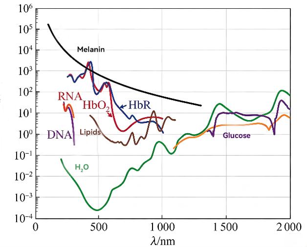
(Absorption-spectrum diagram of selected human-body constituents)
2.2 Pulse-width Requirements for Photoacoustic Imaging
Pulse-width selection is mainly restricted by thermal-constraint conditions. To generate efficient photoacoustic signals, laser pulse width must be far shorter than the thermal-diffusion time of tissue absorbers. Pulse widths within hundreds of nanoseconds are generally required; in practice, the range of 1-100 ns is widely used, among which 4-20 ns is the most common interval. Narrower pulses (~2 ns) can excite photoacoustic signals with broader frequency bands, which theoretically improves axial resolution, yet impose higher requirements on laser design and Q-switching technology and raise costs accordingly.
2.3 Repetition-frequency Requirements for Photoacoustic Imaging
Repetition frequency determines imaging frame rate and data-acquisition efficiency. For deep-tissue-detection-oriented photoacoustic tomography, low-repetition-frequency lasers of 1-10 Hz are typically deployed to guarantee sufficiently high single-pulse energy for penetrating tissue several centimeters thick, at the cost of slow imaging speed. For photoacoustic microscopes, kilohertz-range high-repetition-frequency light sources are frequently employed to realize fast point-by-point scanning.
2.4 Energy Requirements for Photoacoustic Imaging
Pulse energy governs signal strength and maximum imaging depth. In deep-tissue imaging, light attenuates when propagating through tissue before reaching target regions; hence millijoule-level single-pulse energy output from lasers is required. Reported systems feature energies ranging from tens of millijoules up to the joule level. For superficial high-resolution imaging that does not require deep penetration, required energy can drop drastically down to microjoule or even nanojoule levels (e.g., 3 μJ suffices for some applications). Increasing pulse energy improves signal-to-noise ratio, yet operations must stay within tissue-safety thresholds; energy cannot be increased without limit.
III. Matching Lasers for Photoacoustic Imaging
With rapid advances in biomedical-imaging technology, photoacoustic imaging, characterized by non-ionizing radiation, high imaging contrast and large tissue-penetration depth, has been extensively applied in basic biological research, clinical disease diagnosis, intraoperative precision navigation and other scenarios. It places high adaptability demands on supporting laser sources: lasers should not only match tissue-absorption properties precisely but also satisfy strict specifications for output-energy stability, pulse width, parameter-adjustment range, beam quality and other core performances.
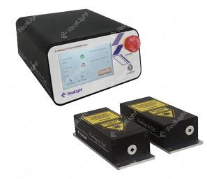
To meet differentiated test requirements for various biological tissues, detection depths, imaging accuracy and dynamic monitoring in photoacoustic imaging, RealLight has independently developed three laser-series: MCA, MCC and MCO. These deliver full-category photoacoustic-imaging laser-source solutions covering narrow pulse width, high repetition frequency, multi-wavelength and high energy. They fully support photoacoustic-signal excitation and precise imaging detection for diverse biological tissues, and offer both free-space output and fiber-coupled output modes for system integration in multiple scenarios.
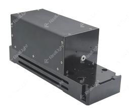
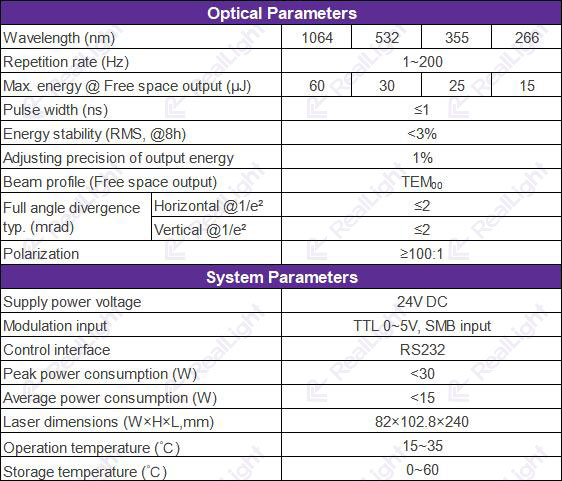
(Physical picture of MCO-series laser; specification table of MCO-series laser)
The three series feature distinct technical advantages in core performance and parameter configuration to satisfy diversified demands of mainstream photoacoustic-imaging equipment.
· MCO series lasers: Offer broad wavelength coverage including four mainstream wavelengths: 1064 nm, 532 nm, 355 nm and 266 nm, precisely matching differential optical-absorption characteristics of different biological tissues and biomolecules for multi-dimensional imaging. Energy can be accurately tuned according to imaging depth and tissue light-transmittance: 1064 nm for deep-tissue biological imaging with continuously adjustable energy from 1 μJ-60 μJ; 532 nm for high-precision fine superficial-tissue imaging with energy adjustable from 1 μJ-30 μJ. Fine energy adjustment is also available for 355 nm and 266 nm ultraviolet bands to satisfy experiments such as special-biological-sample detection and molecular-targeted imaging. This series supports wide-range repetition-frequency tuning from 1-200 Hz, accommodating both low-speed static-tissue scanning and high-speed biological dynamic imaging to capture real-time dynamic changes in tissues and blood vessels.
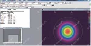
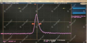
(Beam profile; typical pulse waveform)
· MCA-series sub-nanosecond microchip lasers: Highlight narrow pulse width and low thermal interference, with minimum pulse width down to 1.2 ns. They effectively suppress thermal-diffusion artifacts in imaging and greatly boost photoacoustic-imaging resolution and detection accuracy. Maximum single-pulse energy reaches 120 μJ. Parameters can be flexibly adjusted to balance imaging efficiency and quality. Adopting standard TEM₀₀ fundamental-mode output, they deliver excellent beam quality and high polarization stability. Featuring fully-sealed integrated construction, compact footprint and low power consumption, they suit high-precision, high-stability static fine imaging and long-term experimental measurement scenarios.
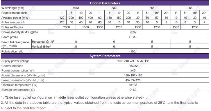
(Physical picture of MCA-series laser; specification table of MCA-series laser)
· MCC-series microchip lasers: Preferred solutions for high-speed dynamic photoacoustic imaging. Pulse width is as low as 750 ps with pure pulse waveform and outstanding output stability. Thermal effects are maximally suppressed for ultra-high-precision, ultra-fast-response dynamic imaging. Maximum repetition frequency reaches 30 kHz for faster response to accurately capture instantaneous dynamic changes in biological tissue. They are ideal for cutting-edge scenarios including in-vivo dynamic imaging and microcirculatory blood-flow monitoring. Output power ranges from 4 mW-400 mW with strong parameter adjustability. Compact and highly reliable, they are compatible with miniaturized imaging-system integration.
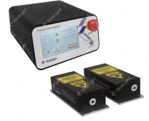
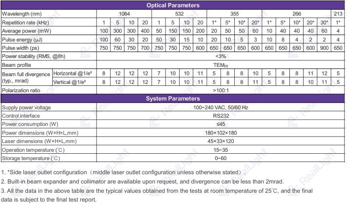
(Physical picture of MCC-series laser; specification table of MCC-series laser)
For deeper-imaging-depth applications, RealLight also supplies high-energy solid-state lasers. HQF-series lamp-pumped Q-switched nanosecond lasers: Compact nanosecond lasers adopting single-lamp-double-rod configuration. Flashlamps feature long service life and easy replacement. Output beams possess flat-top intensity distribution. Single-wavelength or dual-wavelength variants are available. At 1064 nm, output energy can reach 800 mJ and 1.2 J respectively. This series features compact structure, high stability, excellent beam quality and high-energy output.
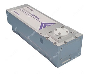
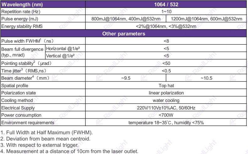
(Physical picture of HQF lamp-pumped Q-switched nanosecond laser; specification table of HQF)
To facilitate integration of high-energy lasers into other equipment, RealLight provides AQE-series RealShock® diode-stack-pumped actively Q-switched lasers. Independently-developed semiconductor stacks realize multi-directional uniform pumping for high-energy, high-stability actively Q-switched laser output with outstanding beam uniformity. Laser resonators adopt hermetic packaging with integrated drive-and-control circuits.
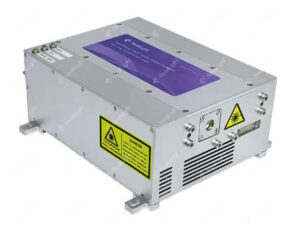
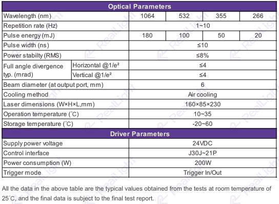
(Physical picture of AQE-180 mJ laser; specification table of AQE-180 mJ laser)
Disclaimer: These are research-grade lasers intended exclusively for laboratory scientific research. They are not medical devices and shall not be directly used for human clinical diagnosis or therapy. References herein to clinical-application directions of photoacoustic imaging represent merely technical-development reviews and do not signify that these products possess clinical-medical functions. Portions of this document are sourced online for technical-research-exchange and reference-and-learning purposes. Please notify us for verification and removal should there be factual errors, academic inaccuracies or copyright concerns.
Referências
[1] Alexander Graham Bell. On the production and reproduction of sound by light[J]. Sciences, 1880, 17(20):305-324.
[2] Shen Xiaowen, Wu Hongpeng, Dong Lei. Research progress of photoacoustic-imaging technology in biomedicine (Invited)[J]. Acta Photonica Sinica, 2023, 52(03):0352105.
[3] Yao J, Wang L V. Functional photoacoustic microscopy for high-resolution and noninvasive in vivo imaging[J]. Laser & Photonics Reviews, 2013, 7(6):758-778.
Q&A
Q: What are the main factors to consider when selecting laser-excitation wavelengths for photoacoustic imaging? What scenarios are suited for typical individual wavelengths?
A: Wavelength selection is primarily determined by target-absorption characteristics and imaging-depth requirements.
· 1064 nm (NIR-II window): Low tissue scattering; suitable for deep large-blood-vessel and contrast-agent imaging. Drawback: strong water absorption.
· 532 nm: Hemoglobin-absorption peak with high photoacoustic-conversion efficiency. Suitable for superficial high-resolution microscopic imaging, yet penetration is limited by scattering.
· 808 nm: Matches the absorption peak of ICG contrast agents, widely used in targeted molecular imaging.
· 785 nm: Used to differentiate oxy-hemoglobin and deoxy-hemoglobin for multispectral photoacoustic imaging.
· Ultraviolet 355 nm / 266 nm: Enable label-free cell-nucleus imaging, yet penetration is only at the micrometer scale with high photodamage risk.
Q: What core advantages does photoacoustic imaging possess compared with conventional optical and ultrasound imaging?
A: Photoacoustic imaging combines merits of both optical and ultrasound imaging.
1. High contrast: Imaging relies on light-absorption differences among different tissue components. Functional imaging can resolve blood-oxygen saturation and reflect tissue metabolism and compositional information.
2. Large penetration depth: Surpasses the ~1 mm depth limit of conventional optical imaging; some systems achieve imaging depth up to 70 mm.
3. Wide resolution range: Spatial resolution spans from micrometers to millimeters.
4. Non-invasive and non-ionizing detection: No ionizing radiation.
5. Multi-scale imaging: Supports imaging from sub-cellular and cellular scales to tissue-and-organ scales, and acquires multi-dimensional information covering anatomy, physiology and molecules.





 777